
Natural History Illustration of a Chironomid Midge

I recently was commissioned to complete a natural history entomological illustration for an expert in fossilized midges at the Natural History Museum in London, as a surprise retirement present.
Studying Chironomid midges
The recipient is Steve Brooks, who examines the fossilized heads of non-biting midges (Chironomids) to examine climate change through time. For an overview of his work, do take the time to read this article, it’s fascinating.
The person commissioning the work (Kimberley Davies) not only is an expert in midges herself, but also grew up in Hay-on-Wye (where I’m based). An extra incentive to make sure I did a good job!
To give you a taste of what I had in store, here is a photo of one of these little insects.
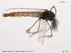
Chironomid midge Micropsectra radialis adult, (photo copyright NTNU Museum, Norway)
Getting good reference for a tricky job
I should point out that although I have a Zoology degree, I am by no means an expert in the world of flies (or Diptera). This job was an incredibly steep (and thrilling) learning curve.
Kimberley was very helpful, providing me with realms of reference images and papers. We decided the best way to construct the illustration would be to have an adult midge, a larval midge, and then some much more detailed illustrations of the head parts below.
Fortunately there are some very good scientific websites out there. This includes one which has lots of photographs of the adult midges and specimens from similar families, BOLD systems taxonomy. Here I found a rich seam of photos of my particular midge, Micropsectra radialis. Most were from the NTNU Museum of Natural History and Archeology in Norway.
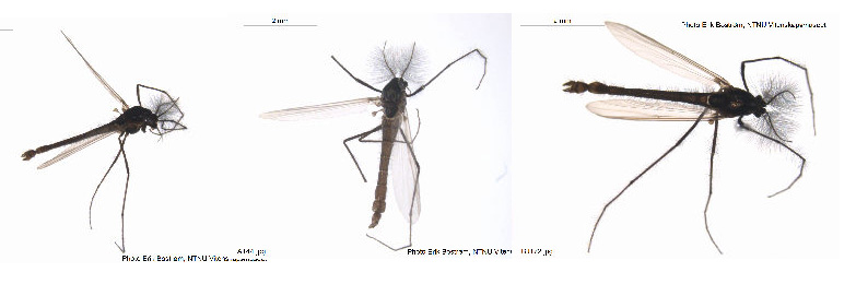
Photos of adult M. radialis specimens, (photos copyright NTNU Museum, Norway)
Armed with this reference, and having found a lovely page of reference of the characteristics of this family of flies, I drew up the adult midge and the larval form.
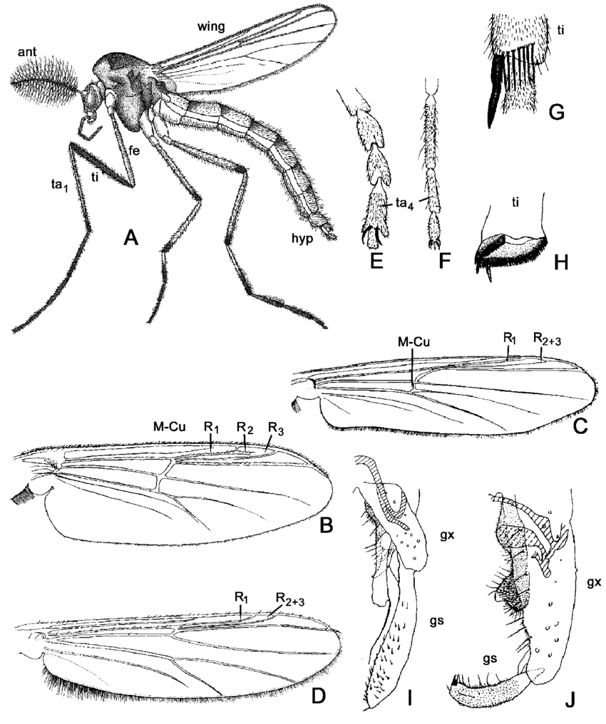
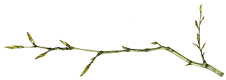
Chironomid midge diagram, From “Chironomid” article on reaearch gate by Peter Cranston.
Illustrating the midge larvae
The larvae look pretty similar to one another unless they’re dissected out under the microscope, they tend to be white or cream with dark head capsules. We were both happy for me to go ahead and add colour to the larva.
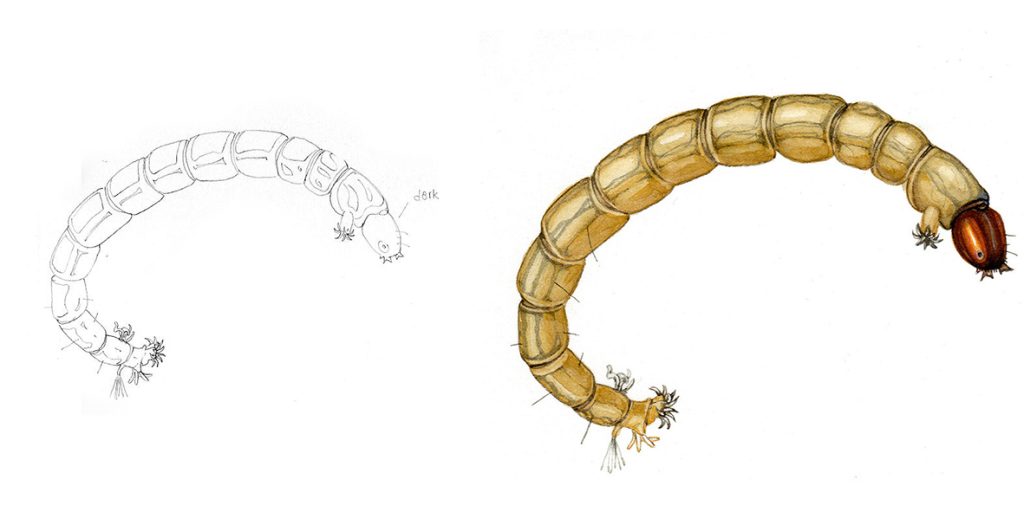
Larva of M. radialis, pencil rough and completed watercolour.
Illustrating the midge adult
Next up was the adult midge, these are about 2mm long. Illustrating this little insect was tricky as the photographs I was working from weren’t great on detail. I had to combine diagram info with photo details and try and get the colours right. The thorax details were particularly difficult to untangle and illustrate. Wings and legs were simple; wings from a diagram and legs from the photo reference. One of the most marvellous features of this species is the males have wonderfully flamboyant antennae, thick and plumed. These were an absolute treat to draw and paint.
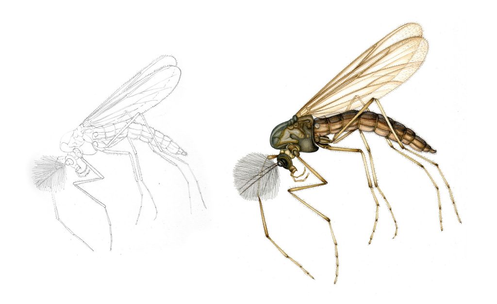
M. radialis adult male, pencil rough and final watercolour
Researching the midge head capsule
Having completed the adult and larvae, we moved onto the details which are species specific. These had to be assembled from lots of photos of microscopic fossilized specimens. Here is some of the reference Kim supplied. This is a page of photos of the microscopic mouth parts of the larval stages of various closely related midge species. The species we were illustrating is Micropsectra radialis. It’s mouth parts are at letter “D”.
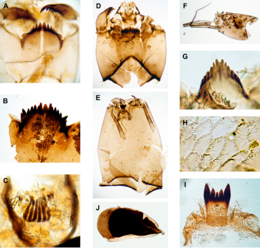
Head capsule details of Chironomid midge larvae (reference from NHM London)
My first attempts at the head capsule details were a touch sketchy, so Kim provided a couple of annotated anatomical diagrams to work with. This made matters far easier.
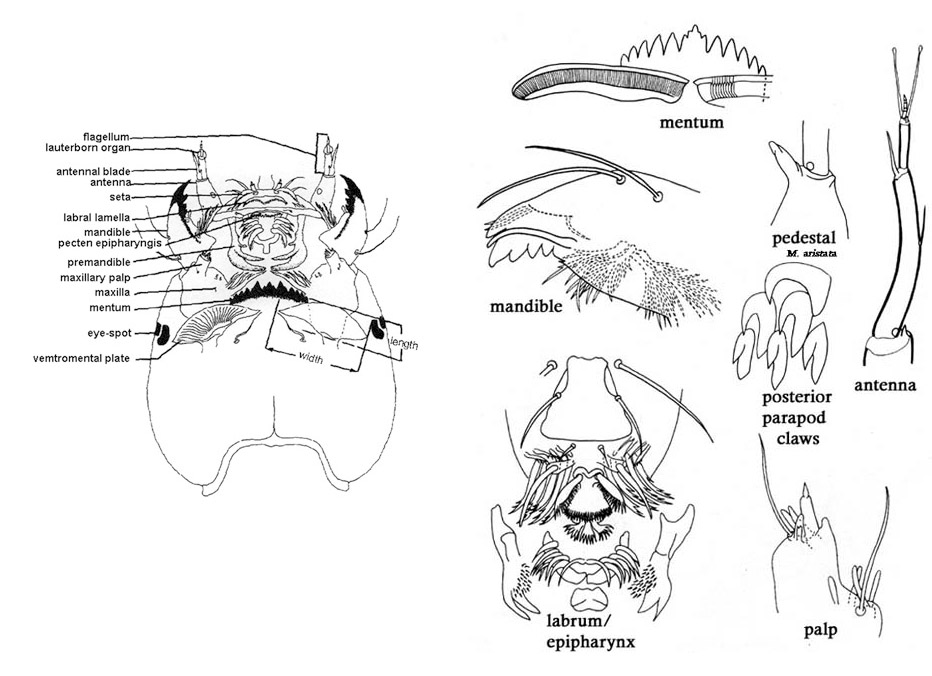
Reference of the head caspsules of M. radialis, provided by NHM London
Kim had enormous patience. I learned terms such as mentum, antenennal pedestals, and vemtromental plates. These were then illustrated. We finally got an illustration of the relvant details thrashed out. Below is a series of the roughs with the earliest being at top and the final version at the bottom.
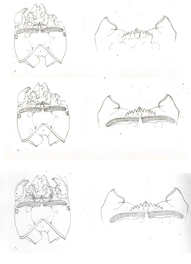
Head capsule details of M. radialis, series of roughs
Illustrating the midge head capsule
Once these details were given the go-ahead, I had to figure out how to add colour. I finally settled on an ochre-pink shade, echoing the colour of the specimens. Where areas of the head capsule overlapped, the shade would be a little darker (like two sheets of coloured cellophane being darker where they overlay each other). The one area of these that I knew was vital was the mentum or teeth. They were sharp and dark with a pale space in the centre of the array. This was easy to accentuate.
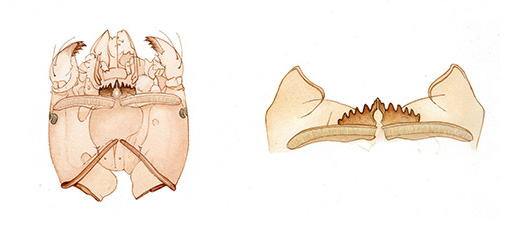
Coloured version of the M. radialis head capsule detail (4th instar) with mentun accentuated
Despite having had reservations, I was pleased with the end result of these details. I was also relieved that the illustration worked out as I was working on a paper which is comparatively new to me. This is hot press watercolour paper, Moulin du Roy, which takes detail very well but is less able to handle layers of watercolour wash. (For more on testing papers, see my blog)
So the painting was complete. Luckily both Kim and Steve Brooks seemed pleased.
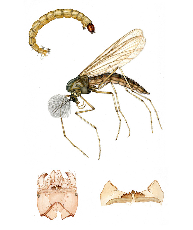
Chironmid M. radialis final illustration
I absolutely loved working on this project, I adore being pushed into very specific corners of the natural world and having to understand and interpret them, and being expected to comprehend all the minutae and species specific information being given to me. It reminds me of being at college, and almost feeling your brain expand as new information pours in, and is yet another reason why I absolutely love my chosen career.
I loved your drawings. Are they available for use in a scientific publication (a chironomid manuscript on which we want to illustrate a method. Please reply to jeffram@wayne.edu (Wayne State University)
Hi Jeffrey
Im about to email a reply to you back, but in theory yes! Thanks.
Lizzie